Please ensure Javascript is enabled for purposes of website accessibility

Dry Eye Options in New Jersey

What is Dry Eye?

Dry eye happens when your eyes don’t make enough tears to stay wet, or when your tears don’t work correctly. This can make your eyes feel uncomfortable, and in some cases it can also cause vision problems.

Dry eye is common — it affects millions of Americans every year. The good news is that if you have dry eye, there are lots of things you can do to keep your eyes healthy and stay comfortable.

Symptoms of Dry Eyes

Dry eye syndrome can cause a variety of symptoms, including scratchiness, stinging, or burning in the eyes. You might also experience redness, eye sensitivity, blurred vision or feel as if there is something in your eye. If you’re experiencing any of these symptoms, it’s important to see your eye doctor as soon as possible. Your symptoms can get worse over time if left untreated, or can even develop into more serious eye conditions and result in corneal damage.

Who is at Risk for Dry Eyes?

While dry eyes can affect everyone, you might be more at risk if you’re over the age of fifty, are female, live in a dry climate, wear contacts, or have a Vitamin A deficiency. Autoimmune conditions such as lupus, Sjogren syndrome, and arthritis, among others, can also make a person more susceptible to dry eyes. Jobs that involve long hours of staring at a computer or tablet screen can also contribute to dry eyes.

How is Dry Eye Syndrome Diagnosed?

A comprehensive dilated eye exam is the best way for your doctor to determine the underlying cause of your dry eyes. This includes a complete history of your overall health which can help yput eye care specialist diagnose the cause of your dry eyes.

In addition to a vision test, your doctor can perform tests to see how many tears you produce, look at the structure of your eyelids and glands, and check how long it takes for your tears to dry. This test will include pupil dilation, in which your provider will put drops into your eyes to widen the pupils.

Treatments for Dry Eyes

Many cases of dry eyes are due to blocked oil glands, but other causes are low tear production, tears that dry too quickly, and tears that drain too quickly. In most cases, medication or eye drops can be prescribed to address the issue and alleviate symptoms.

Depending on the severity of the dryness, you might also be prescribed pills, eye drops, nasal sprays, or even eye inserts to help you produce more tears. Some of these products are prescription strength, but for mild cases, your doctor can prescribe over-the-counter eye drops also called artificial tears..

If tears drain too quickly from your eyes, your doctor may suggest plugs for your tear ducts. In severe cases of tear drainage, a doctor might suggest surgery to tighten eyelids, but this treatment is rare.

Schedule A Consultation

If you’re experiencing any of the above symptoms, please reach out to us and schedule a consultation with one of our skilled providers at Campus Eye Group in Hamilton, NJ. We offer comprehensive eye services, including diagnostic and treatment options for dry eyes. Call us at (609) 587-2020 or use our online contact form.


Risks of not taking care of your cataracts

Cataracts are a degenerative condition that affects the lenses in one or both eyes, creating a cloudiness that makes it harder to see as the condition progresses. For whatever reason, some individuals may hold off on getting the treatment they need for their cataracts. But is that a wise decision? If you have cataracts and you’re potentially putting off treatment as the condition progresses, let’s take a closer look at the risks of not taking care of your cataracts and how you can get the support you need today with Campus Eye Group.

Your Lifestyle Will Be Greatly Impacted

Cataracts, even mild ones, have an impact on your vision. For those who see just fine, this isn’t something they think about often. But once your vision starts to worsen, it makes navigating your environment or doing something as simple as driving around impossible. It can make it difficult to carry out the basic duties of your job, prevent you from seeing friends and family, and complicate your life in other ways. Put simply, if you don’t take care of your cataracts, you risk changing your life in many impactful ways.

Your Vision Could Become Seriously Impaired

You might be holding off on having cataract treatment because you’re worried about costs or because your cataracts haven’t progressed to a point where you’re experiencing major side effects as a result. The problem? Even if things don’t seem bad now, you run the risk of letting your vision worsen to the point where you’re unable to do anything about it. After a certain amount of time, cataracts that have progressed too far pose a substantial surgery risk. This means that you won’t be able to get back your vision like you would if you had taken care of the issue early on.

Your Case Could Progress Rapidly Despite Mild Symptoms Now

Some people can manage mild symptoms of cataracts with glasses, making it so that the side effects are tolerable. They think that, because it’s mild now, they have time to correct it later. The decline of your vision could be rapid. While things don’t seem bad now, that’s not indicative that your vision will be a slow decline, giving you plenty of time to remedy it later. The longer you wait, the riskier your condition becomes, and it may progress much faster than you believe it could.

Schedule a Consultation

The risks of leaving cataracts untreated are myriad, which is why it’s so important to seek out treatment the moment you realize you need help. If you’re seeking help with your cataracts in Hamilton, NJ, start here with Campus Eye Group! We are a leader in eye care and are dedicated to making sure you get the proper treatment for your cataracts, especially if you’re in the early stages of the condition and ready to make a change that bolsters your vision and supports your future. If you’re ready to improve your eye health, contact us today to schedule your eye exam.

How to choose your LASIK Surgeon

Choosing the right surgeon for your LASIK procedure is critical, as the results of LASIK surgery will significantly impact your vision and overall quality of life. There are important considerations that should guide your choice of a LASIK surgeon to ensure the best possible outcome.

Qualifications and Experience

All surgeons performing the LASIK procedure should be board-certified in ophthalmology, hold the necessary licenses to perform LASIK, and have updated certifications. You should check where your surgeon received their education and training. Prestigious universities indicate a top-notch education, and the better the education and training, the better the doctor is at performing the procedure and caring for patients.

In addition, the number of LASIK surgeries performed is a significant consideration. It is essential to ask about the surgeon’s experience and the number of surgeries performed. It takes at least ten years to develop the breadth of knowledge required to comfortably complete the LASIK procedure, as this provides sufficient practice with a wide range of cases and unique situations. It is not unusual for an experienced surgeon to have performed more than 25,000 LASIK surgeries.

Reputation

Ensure the surgeon you are considering has an impeccable reputation with former patients and professional associations such as the American Academy of Ophthalmology. Reading patient reviews and testimonials can provide insights into whether others were satisfied. You can request additional references if you would like a more personalized referral. Former patients must have consistent positive feedback.

Technology and Equipment

The LASIK center should invest in the latest technology and equipment. Ask your LASIK experts about the equipment they use and why they have chosen it. Also, the LASIK center should adhere to strict hygiene standards, safety protocols, and infection control measures.

Consultation and Evaluation

A thorough examination is necessary to determine whether you are a suitable candidate for LASIK. Your LASIK center experts should conduct a comprehensive eye examination, discuss your medical history, and address any concerns and questions you may have. They should also provide realistic expectations regarding the outcome of the procedure.

Also, the LASIK Center should provide comprehensive follow-up care after the procedure and be available afterward should any complications or concerns arise.

Communication and Trust

A good LASIK consultant will care enough to take the time to answer any questions you may have, explain the procedure in detail, and make sure that you feel comfortable and informed during the process. Trust and open communication are paramount to achieving a satisfactory result.

Finally, while cost should not be the sole factor in determining your LASIK provider, you should understand the pricing structure and financing options.

Schedule a Consultation

Campus Eye Group is committed to providing patients with superior outcomes in a safe and comfortable environment. To find out if you are a good candidate for LASIK surgery, schedule a consultation with one of our board-certified ophthalmologists and expert LASIK surgeons in Hamilton, NJ, today.

How to Identify and Treat Your Eye Condition

Our eyes are a vital part of our day-to-day life. As we age, our eyes are exposed to multiple health risks. These health risks may lead to the deterioration of your sight or even total blindness. Though small, the eye is a mighty organ with a multitude of working parts, but with so many different parts, self-identifying your condition is nearly impossible. Luckily, you are not alone.

With our expert staff of board-certified ophthalmologists and optometrists, Campus Eye Group offers a comprehensive range of services, from regular eye exams to the treatment of eye conditions, such as:

These are just a few examples– Campus Eye Group is a full-service facility treating all eye conditions, including complications from wearing contact lenses. Our expert staff can help you identify eye conditions and come up with a treatment plan that best suits your needs. Below is a list of the most common eye conditions we see and treat:

Glaucoma

This is a serious eye condition in which there is too much pressure from within the eye. Over time, this pressure damages the optic nerve. Sight loss and even total blindness are possible. Fortunately, glaucoma can be treated if caught early.

Glaucoma typically doesn’t have warning signs until significant eye damage has occurred. This, amongst a long list of other reasons, is why routine eye exams are so important. Your eye care professional will measure your internal eye pressure at every routine eye exam, which can help to catch glaucoma before any damage occurs.

Diabetic Eye Disease

When diabetes is untreated or poorly controlled, high blood sugar levels can damage critical blood vessels in the eye’s back surface called the retina. Your eye doctor can see this damage during an eye exam. In fact, some people may first discover they have the disease in this way. Like glaucoma, diabetic eye disease is often silent until irreparable damage has been done. Regular eye exams are your best protection, especially if you already know you have diabetes.

Macular Degeneration

This disease primarily affects older individuals, most often past the age of 50. Smokers and those with a family history of macular degeneration are more prone to the condition. Macular degeneration causes the center of the retina, called the macula, to break down or grow extra blood vessels underneath it. These vessels break and bleed, damaging the macula. Symptoms include fuzzy images in the center of your vision and lines of letters or numbers that jump or appear to be uneven when they aren’t. Like every eye condition, a professional diagnosis and treatment plan is necessary for the health of your vision.

Conjunctivitis

Commonly called pinkeye, conjunctivitis has many causes, including eye injury, allergies, reaction to certain chemicals, and infection. Symptoms include a pink or red appearance to the white part of the eye, sensitivity to light, pain, watering, and eye discharge. Conjunctivitis is usually self-identifiable and isn’t always serious. However, only an eye care professional can determine the correct cause and treatment.

LASIK

This procedure uses a special laser to reshape the cornea, eliminating or reducing far and near-sightedness and improving astigmatism, which is an irregular curvature of points in the eyeball. LASIK may restore 20/20 vision for some individuals. Understanding when LASIK is necessary and undergoing the surgery can only be done with the assistance of board-certified professionals. Results are generally permanent and may eliminate the need for glasses and contacts altogether.

Campus Eye Group

Our practice is proud to serve the Hamilton, NJ, and Princeton, NJ areas. We warmly welcome new patients, and invite you to call our office at (609) 587-2020 with questions. Our staff is highly qualified and is available at your convenience to perform eye exams and treatment plans. Schedule an appointment to ensure your vision’s health at Campus Eye Group.

Who is at Risk for Cataracts?

Has your vision been a little blurry lately? If so, the problem might be a cataract.

Of course, you’ve probably heard the word “cataract” many times before. But what exactly does it mean?

When the proteins that form your eye’s lens start to break down and stick together, cloudy areas called cataracts can form.

Cataracts can be scary, and if left untreated, they can lead to permanent blindness.

Fortunately, routine eye exams, cataract removal surgeries, and lens replacement operations can all save and protect your eyesight.

At this point, though, you might be wondering: Who is most likely to suffer from this problem?

To answer that question, let’s look at some of the major cataract risk factors.

Age

A person’s age is the primary contributing factor to cataracts. In fact, the proteins of the lens start to break down around the age of 40. And symptoms of cataracts typically start to appear after a person turns 60.

Gender

Women run a somewhat higher risk of cataracts than men, especially earlier in life. It’s not precisely clear why, but it may be connected to the hormonal changes that women can experience, particularly during pregnancy and menopause.

Diabetes

When people have diabetes, their higher blood sugar levels can damage their ocular blood vessels, making them more vulnerable to cataracts. On top of that, the extra blood sugar can cause structural changes within the lens itself. And those changes can lead to cataracts.

Smoking and Drinking

Cigarette smoke can irritate the eyes and swell their blood vessels, which can induce cataract formation.

Moreover, medical studies have linked ongoing heavy alcohol consumption to cataracts. Heavy drinking is often defined as eight or more drinks in one week for a woman and 15 or more drinks in one week for a man. However, the connection between alcohol and cataracts still needs further study.

Genetics

In some cases, cataracts can run in families. A parent might pass on the genetic mutation that causes cataracts to their child.

Other Eye Problems

Those who’ve experienced eye-related traumas, disorders, or injuries are often more susceptible to cataracts. All of these issues can alter the structure of the lens and its proteins.

Schedule A Consultation

Whether or not you’re experiencing cataract symptoms, regular check-ups are vital to the health and functioning of your eyes. The earlier a cataract or other problem is detected, the better the outcomes are likely to be. That’s where we come in. At Campus Eye Group. our experienced team of specialists can help you maintain your eye health. Simply call our office today at (609) 587-2020, or use our online contact form to schedule a consultation.

What is Macular Degeneration?

Macular degeneration is a progressive eye disease affecting the macula, the central part of the retina responsible for sharp vision. It is a leading cause of vision loss in people over 50. But how do you know if you have it? There are some signs and symptoms you can look out for, as well as some ways to prevent it from happening in the first place.

Signs and Symptoms of Macular Degeneration

Blurred or Distorted Vision

One of the most common early symptoms of macular degeneration is blurry or wavy central vision. Straight lines may appear distorted, and objects may lose their clarity.

Difficulty Reading or Performing Close-Up Tasks

As the condition progresses, individuals may find it increasingly challenging to read small print, sew, or perform other activities that require detailed or more precise vision.

Dark or Empty Areas in Your Central Vision

Some people with macular degeneration may experience dark or empty spots in their central vision. This can make it difficult to recognize faces, drive, or engage in activities that require focused vision.

Decreased Color Perception

Macular degeneration can cause a decrease in color vibrancy or intensity. Colors may appear faded or less vivid than before.

Difficulty Adapting to Low-Light Conditions

Individuals with macular degeneration may find it harder to adjust to dim lighting or require brighter lighting to see things more clearly.

Gradual Loss of Central Vision

Macular degeneration typically affects central vision first, while peripheral vision remains intact. Over time, the loss of central vision can become more pronounced and impact daily activities.

Types of Macular Degeneration

Dry Macular Degeneration

This is the more common type, accounting for about 90% of cases of macular degeneration. It is characterized by the gradual breakdown of light-sensitive cells in the macula. The progression of dry macular degeneration is generally slower than the wet type.

Wet Macular Degeneration

This type is less common but more severe. It occurs when abnormal blood vessels grow beneath the retina and leak fluid or blood, leading to rapid and significant damage to the macula. Wet macular degeneration can cause sudden and severe vision loss.

Best Ways to Reduce Your Risk of Macular Degeneration

Because macular degeneration can be so serious, knowing ways to reduce your risk can be vital to keeping your vision intact. Some of the best ways to reduce your risk include:

  • Consume a healthy diet rich in vitamins, omega-3 fatty acids, and antioxidants. Eat leafy green vegetables, seafood, nuts, carrots, and vibrant fruits to support your eye health.
  • Give up smoking. Smoking significantly increases the risk of macular degeneration. If you already have the disease, quitting smoking can help slow its course.
  • Use sunglasses that block UV rays, and think about using blue light filters on your digital devices to lessen the risk of macula deterioration.
  • Regular exercise: Maintaining general health through physical activity may help improve eye health as well.
  • Routine eye exams: To effectively manage macular degeneration, schedule routine visits with an eye care expert for thorough eye examinations.

Schedule A Consultation Today

As early management can help slow the advancement of macular degeneration and preserve your vision, early identification and routine eye exams are essential in recognizing and treating macular degeneration.

For an accurate diagnosis and proper management, schedule an appointment with Campus Eye Group today. Call our Hamilton, NJ office at (609) 587-2020 or use our online contact form.

What to do before LASIK surgery?

Are you considering LASIK surgery to correct your vision? Before you get LASIK, there are some important steps you can take to make sure you have the best possible experience.

At Campus Eye Group, we want to ensure you have all the information you need to make the best decisions about your vision care. With that in mind, let’s take a look at some of the things you should do before getting LASIK.

Share Your Medical History

Before undergoing any kind of surgery, being honest about your medical history is important. LASIK is a very safe procedure, but your surgeon needs to know about any existing medical conditions and medications you may be taking to assess your candidacy properly. Discuss any eye diseases, autoimmune disorders, pregnancy, or other medical issues with your doctor.

Drink More Water Before Surgery

It’s important to stay hydrated before and after your LASIK surgery. Be sure to drink plenty of water the week before the surgery and during your recovery. Staying hydrated will help your body heal more quickly and reduce the risk of any post-surgery complications.

Stop Wearing Contact Lenses

If you wear contact lenses, stop wearing them at least two weeks before your surgery. Contact lenses can alter the shape of your cornea, so it’s important to give your eyes time to return to their natural shape before your procedure.

Let Someone Drive You to the Procedure

Ensuring you have someone to drive you to and from your LASIK procedure is important. Driving yourself home after the surgery is not recommended as your vision may be blurry, and you may be more sensitive to light. Make sure to arrange for a friend or family member to be your designated driver for the day.

Don’t Wear Makeup

It is important to avoid wearing makeup, on the day of your LASIK procedure. Makeup can irritate your eyes after the surgery and increase the risk of infection.

In addition, it’s important to wash your face with a mild cleanser the night before the procedure, as any makeup residues can also increase your risk of infection. Be sure to arrive at your appointment with a clean face.

Wear Comfortable Clothes

It’s important to wear comfortable clothing to your LASIK appointment. Avoid wearing tight-fitting clothing as it could affect your eyes when you’re trying to take it off. Wear a shirt or blouse that buttons up in the front or that has a wider neckline.

Be Mentally Prepared

LASIK is a surgical procedure, so being mentally prepared for the experience is important. Talk to your doctor about what to expect during the procedure, and ask any questions you may have. Knowing what to expect can help you relax and stay calm during the procedure.

Bring Sunglasses

It’s also important to bring sunglasses to your LASIK appointment. Your eyes will likely be more sensitive to light right after the surgery. To protect your eyes from the light and UV rays and to stop you from rubbing or scratching your eyes, sunglasses are recommended. You can also bring a hat to wear to block out the UV rays and light that may make it over your sunglasses as well.

Learn About Post-Surgery Care

Before your procedure, your doctor will provide detailed instructions on how to care for your eyes after the surgery. Be sure to follow these instructions and avoid any activities that could strain your eyes in the days following the procedure.

Schedule A Consultation

LASIK surgery can be a life-changing experience that can correct your vision and allow you to see clearly without glasses or contacts.  At Campus Eye Group, our experienced team of eye care professionals will be happy to answer any questions and help you prepare for your procedure.

To schedule a consultation today, call our offices or use our online scheduling form.

Types of Cataracts and Symptoms to Look For

Cataracts are a degenerative disorder that causes one or both of your eyes’ lenses to become cloudy. They impair your vision and can lead to blindness if not appropriately addressed. Cataracts can be caused by aging, long-term UV exposure from sunlight, or diabetes.

Cataracts develop slowly over time, and symptoms may only begin to show around age 40 for most people. If you’re beginning to notice cloudiness or unclear vision, it may be cataracts. Below we discuss types of cataracts and what symptoms to look out for.

Types of Cataracts

It is estimated more than 24.4 million Americans aged 40 and older have cataracts. By age 75, almost half of all Americans have some form of visual disability due to cataracts. Although they’re usually not painful, they must be treated because they will gradually affect your eyesight as the condition worsens.

There are four main types of cataracts.

1. Nuclear Cataracts

A nuclear cataract is the most common type of cataract, beginning with a gradual hardening and yellowing of the central zone of the lens also known as the nucleus. Over time, this hardening and yellowing will expand to the other layers of the lens.

As this type of cataract progresses, it changes the eye’s ability to focus and close-up vision (for reading or other types of close work) may temporarily improve. This symptom is referred to as second sight, but the vision improvement it produces is not permanent.

A nuclear sclerotic cataract progresses slowly and may take several years of gradual development before it begins to affect vision.

2. Cortical Cataracts

A cortical cataract forms in the shell layer of the lens known as the cortex and gradually extends its “spokes” from the outside of the lens to the center. These fissures can cause the light that enters the eye to scatter, creating problems with blurred vision, glare, contrast and depth perception. People with diabetes are at risk for developing cortical cataracts.

3. Posterior Subcapsular Cataracts

Primarily affecting one’s reading and night vision, this type of cataract begins as a small opaque or cloudy area on the posterior (back surface) of the lens. It is called subcapsular because it forms beneath the lens capsule which is a small sac or membrane that encloses the lens and holds it in place.

Subcapsular cataracts can interfere with reading and create halo effects and glare around lights. People who use steroids or have diabetes, extreme nearsightedness, and/or retinitis pigmentosa may develop this type of cataract. Subcapsular cataracts can develop rapidly and symptoms can become noticeable within months.

4. Congenital Cataracts

These are rare, but they can also happen to anyone. They’re when your lens develops abnormally before birth or during childhood. The lens of the eye is normally clear.  It focuses light that comes into the eye onto the retina. Cataract develops because the lens is positioned incorrectly.

Symptoms to Look For

Cataracts can have a variety of symptoms depending on their severity. As cataracts develop, you may experience the following:

  • Blurry Vision – the first sign you might see is something out of place in your vision. This could mean the appearance of halos around objects like street lamps, lights, signs, and buildings. You may also notice that your vision suddenly seems darker than before, even in bright light.
  • Double Vision – this occurs when you have a cataract that has simultaneously affected both eyes’ lenses. The image from each eye is often out of alignment, so both images appear upside down or reversed.
  • Increased light and glare sensitivity – your eyes may be more light-sensitive. This could mean you have to shield them from the sun when you go out or turn off any bright lights you have in your home.
  • Limited night vision – it can be difficult to see in the dark with cataracts. You can have trouble driving at night, reading street signs, and identifying objects.
  • Loss of peripheral vision – this usually occurs when your cataract has become severe enough to affect both eyes at once and impair your field of view. Objects that are close by appear to be farther away, or vice versa.

Schedule an Appointment

If you experience these symptoms, Campus Eye Group can help. Our knowledgeable ophthalmologists can determine whether cataracts are impacting your vision and how they can be removed. Contact us today to schedule an appointment.  

Contoura Vision: What to Expect?

Contoura™ Vision – Topography-Guided Laser Vision Correction

The field of laser vision correction is constantly progressing. Since the advent of laser refractive surgery in the 1980s, more and more people across the world have been able to reduce the effects that nearsightedness, farsightedness, and astigmatism have had on their daily lives – and many have completely eliminated their need for corrective eyewear in order to see clearly. The efficacy of these procedures has continued to improve as the technology has advanced, and our providers at Campus Eye Group have established ourselves as leading voices in the development of new techniques.

What is Contoura and What is Unique About it?

The eye has two kinds of axes: the pupillary and visual axes. The pupillary axis passes through the center of the front sight, while the optical axis is the line that passes through the object of interest and fovea. Unlike other laser eye procedures, Contoura works on the visual axis, which results in better vision.  In fact, Contoura when added with LASIK can produce sharper results than LASIK, the most popular laser eye correction surgery.


ContouraTM Vision – Topography-Guided Laser Vision Correction can optimize the surface of the eye’s cornea in such a way that it enhances the quality of vision better than has ever been possible before. One of the unique characteristics of the cornea is that it does not have a smooth surface. Contoura involves your ophthalmologist using a Topolyser to map the irregularities in your cornea and understand 22,000 unique data points. Understanding the individual data points enables your ophthalmologist to adjust your surgery accurately and effectively. Contoura targets the aberrations of the cornea that need to be corrected.


In clinical studies, ContouraTM Vision – Topography-Guided Laser Vision Correction helped 90% of patients see as well or better without glasses than they did with glasses. It can benefit patients in several ways, helping them obtain a better quality of vision and reducing issues with night glare.


Although previous laser vision correction techniques have offered patients incredible precision and accuracy while reshaping the cornea and addressing visual refractive errors, ContouraTM Vision – Topography-Guided Laser Vision Correction takes the treatment even further. Instead of just treating the refractive error, this technology utilizes unparalleled diagnostic capabilities to identify and record imperfections in the curvature of the cornea and the optics of the cornea, combining those measurements with the refractive error to give the eyes a better-focusing surface. The technique goes together with our state-of-the-art Excimer Laser, allowing our experienced eye surgeons to create a truly customized LASIK procedure that can enhance your eyesight with stunning results.

Schedule an Appointment

During your initial LASIK consultation and eye evaluation our LASIK specialists will talk with you about your eligibility and determine if Topography-Guided Laser Vision Correction would be beneficial for your needs.  Call us today at 609-587-2020 or fill out our web form to get started website form to get started.

Latest Advancements in the Treatment for Age Related Macular Degeneration (AMD)

For more than a decade, ophthalmologists have treated wet age-related macular degeneration (AMD) with eye injections given every month or two, and dry AMD with antioxidant vitamins. These treatments were groundbreaking when introduced, offering hope for the first time that this sight-threatening disease could be slowed, and in some cases stopped or even reversed. This revolution is undergoing an intriguing evolution. So, what will the next decade hold for the 11 million Americans with AMD?

In short, the latest research is varied, vibrant, and suggests a future in which ophthalmologists will have more effective options to protect people from slow vision loss caused by AMD. Here’s a rundown of the most promising AMD treatments on the horizon.

New Treatments for Wet AMD

Wet AMD develops when new, abnormal blood vessels grow under the retina. These vessels may leak blood or other fluids, causing scarring of the macula. You lose vision faster with wet AMD than with dry AMD.

About 15 years ago, scientists created drugs that interfere with this process by blocking a protein called vascular endothelial growth factor (VEGF). Before the creation of these so-called anti-VEGF drugs, people with wet AMD were almost certain to develop severe vision loss or blindness.

Then, in 2005, anti-VEGF drugs broke ground by saving the sight of patients with wet AMD. These drugs stabilize or improve vision in the vast majority of patients. But they must be injected into the eye on a regular basis.

While clinical trials show that anti-VEGF injections have allowed more than 90% of patients to keep their vision, in the real world the percentage is closer to 50%. That’s because people aren’t being treated as regularly as they should. The problem is most people need an injection every four to eight weeks to keep their vision. This can be a difficult schedule to maintain for many elderly patients struggling with other maladies and reliant on others to get them to their ophthalmology visits.

Some of the most exciting research today is looking at better alternatives to frequent injections. It’s not just about convenience; the hope is that a more consistent treatment will also help people keep more of their vision.

Gene Therapy for Wet AMD

Gene therapy is a promising alternative to ongoing eye injections of drugs such as Eyelea, Lucentis and Avastin. The goal of gene therapy is to provide a ‘one-and-done’ treatment by helping the eye make its own anti-VEGF medicine. Two different methods are under investigation: One injects the gene therapy underneath the retina in a surgical procedure; the other injects it into the eye just like a routine anti-VEGF treatment is done in the doctor’s office.

There are four different drug candidates under investigation for wet AMD and one for dry AMD. Despite the promise of gene therapy, the long-term effectiveness remains to be seen. Among the challenges it faces is the likely sky-high cost of such a treatment.

New Methods of Delivering Drugs to the Eye

One promising approach that could be available soon is a refillable drug reservoir. The port delivery system (PDS) is a tiny refillable device that stores the anti-VEGF drug Lucentis. No bigger than a grain of rice, the port is implanted into the wall of the eye, just under the eyelid, during a surgical procedure. The device continuously releases drugs to the back of the eye over time. Instead of an injection every six to eight weeks, patients might get a fill-up once or twice a year at the doctor’s office. The device can be refilled using a special needle. The latest studies show many people treated this way were able to go 15 months between treatments.

Drugs that Treat Multiple Causes of Wet AMD

Anti-VEGF treatments are effective because they target one key factor that contributes to wet AMD: VEGF. But what if one drug could treat two underlying causes of AMD? That’s the idea behind the drug faricimab. It targets both VEGF and the protein angiopoietin-2. It’s injected into the eye like a standard anti-VEGF treatment, but it lasts a long longer. The latest research shows patients could go up to four months in between treatments. However, this data is so new that it has not yet been published in a peer-reviewed journal.

It may also be possible to combine two drugs and hit wet AMD with a double punch. These combos could improve vision and make injections last longer. Cosopt (dorzolamide-timolol), an eye drop already used to treat glaucoma, is being tested in combination with anti-VEGF injections. Studies have shown that the duo can reduce retinal fluid accumulation and last longer than an anti-VEGF injection alone. Opthea’s OPT-302 targets a protein that contributes to wet AMD: angiopoietin II. Combining Opthea with an anti-VEGF injection may work better and last longer than current anti-VEGF injections.

Longer-lasting anti-VEGF injections

Drugmakers are creating new anti-VEGF treatments that do not need to be injected into the eye as often as Eyelea, Lucentis or Avastin.

Novartis’ new drug, Beovu, is now approved for use in the United States. About one in every three patients using Beovu can go for as long as three months before needing another injection. Beovu may do a better job than other drugs of drying retinal fluid in patients with wet AMD.

Allergan’s Abicipar is still in phase 3 clinical trials and has not yet gained FDA approval. Studies suggest this drug allows patients to go three months between injections. This drug is also being evaluated for macular edema.

Another injectable medicine — Sunitinib by Graybug Vision — has the potential to give patients 6 months of durability between treatments. Sunitinab is just entering Phase 2B clinical trials. It may still be 3 to 5 years away from becoming available.

New Treatments for Dry AMD

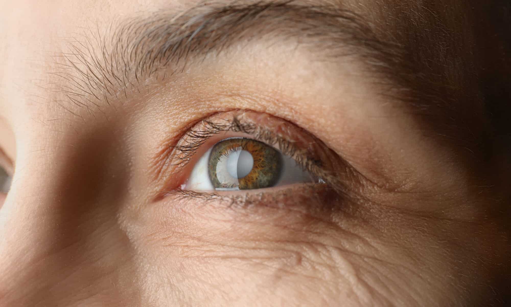
About 8 out of 10 of people with AMD have the dry form. Dry AMD occurs when parts of the macula thin with age, and tiny clumps of protein called drusen grow. You slowly lose central vision. Depending on severity, dry AMD is considered early, intermediate or late stage.

For people with intermediate disease, a formulation of antioxidant vitamins called the AREDS2 formula can help reduce the risk of vision loss. But for people with late-stage AMD, also called geographic atrophy (GA), there is no treatment available. However, there are several promising clinical trials underway.

Dry AMD treatments that target the immune system

A part of the immune system called the “complement cascade” has long been identified as a culprit in AMD. Two new drugs that target the complement cascade and stop it from attacking the retina have recently advanced to late-stage clinical trials. One (pegcetacoplan, APL-2) targets a complement protein called C3, the other drug candidate (Zimura, avacincaptad pegol) targets a different protein in the cascade, C5. Like currently available treatments for wet AMD, these drugs are injected directly into the patient’s eye. Already proven safe in people, researchers are now investigating whether they can substantially improve vision. Results are expected in about a year.

Replacing vision cells in people with dry AMD

Another concept under investigation is the possibility of replacing some cells that begin to die in late-stage dry AMD. Stem cells may be able to replace the retinal cells (light-sensitive cells) that are killed off by this disease. Doctors are devising ways to transplant these stem cells into the eye. One strategy is to layer the stem cells on thin scaffolds. Another tactic is to put the cells into a fluid suspension that can be injected under the retina. Stem cells have been tested in small clinical trials and they do not have unexpected side effects. It may take about 10 to 15 years for these therapies to be fine-tuned and proven effective in humans.

Contact Us Today

Low vision aids like reading glasses and large-print products may temporarily help age-related macular degeneration symptoms, but a long-term solution is essential to prevent total vision loss. Schedule a consultation with an eye doctor at Campus Eye Group today at one of our three New Jersey locations.