Please ensure Javascript is enabled for purposes of website accessibility

How to Identify and Treat Your Eye Condition

Our eyes are a vital part of our day-to-day life. As we age, our eyes are exposed to multiple health risks. These health risks may lead to the deterioration of your sight or even total blindness. Though small, the eye is a mighty organ with a multitude of working parts, but with so many different parts, self-identifying your condition is nearly impossible. Luckily, you are not alone.

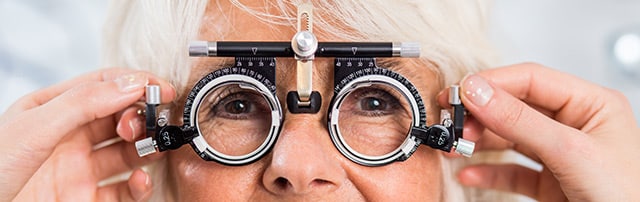
With our expert staff of board-certified ophthalmologists and optometrists, Campus Eye Group offers a comprehensive range of services, from regular eye exams to the treatment of eye conditions, such as:

These are just a few examples– Campus Eye Group is a full-service facility treating all eye conditions, including complications from wearing contact lenses. Our expert staff can help you identify eye conditions and come up with a treatment plan that best suits your needs. Below is a list of the most common eye conditions we see and treat:

Glaucoma

This is a serious eye condition in which there is too much pressure from within the eye. Over time, this pressure damages the optic nerve. Sight loss and even total blindness are possible. Fortunately, glaucoma can be treated if caught early.

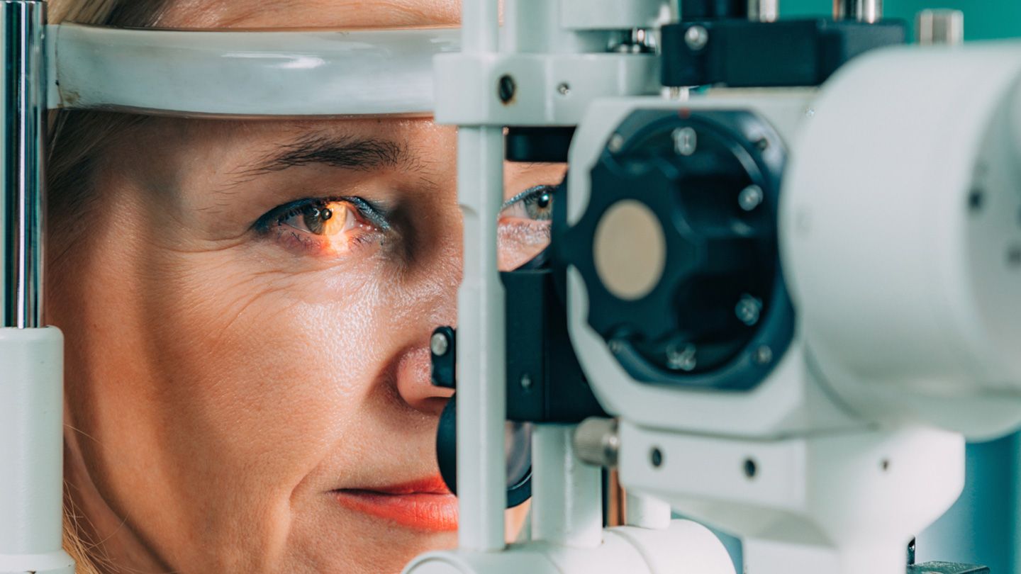
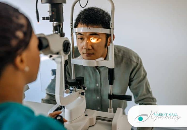
Glaucoma typically doesn’t have warning signs until significant eye damage has occurred. This, amongst a long list of other reasons, is why routine eye exams are so important. Your eye care professional will measure your internal eye pressure at every routine eye exam, which can help to catch glaucoma before any damage occurs.

Diabetic Eye Disease

When diabetes is untreated or poorly controlled, high blood sugar levels can damage critical blood vessels in the eye’s back surface called the retina. Your eye doctor can see this damage during an eye exam. In fact, some people may first discover they have the disease in this way. Like glaucoma, diabetic eye disease is often silent until irreparable damage has been done. Regular eye exams are your best protection, especially if you already know you have diabetes.

Macular Degeneration

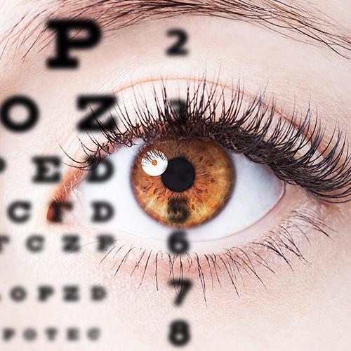
This disease primarily affects older individuals, most often past the age of 50. Smokers and those with a family history of macular degeneration are more prone to the condition. Macular degeneration causes the center of the retina, called the macula, to break down or grow extra blood vessels underneath it. These vessels break and bleed, damaging the macula. Symptoms include fuzzy images in the center of your vision and lines of letters or numbers that jump or appear to be uneven when they aren’t. Like every eye condition, a professional diagnosis and treatment plan is necessary for the health of your vision.

Conjunctivitis

Commonly called pinkeye, conjunctivitis has many causes, including eye injury, allergies, reaction to certain chemicals, and infection. Symptoms include a pink or red appearance to the white part of the eye, sensitivity to light, pain, watering, and eye discharge. Conjunctivitis is usually self-identifiable and isn’t always serious. However, only an eye care professional can determine the correct cause and treatment.

LASIK

This procedure uses a special laser to reshape the cornea, eliminating or reducing far and near-sightedness and improving astigmatism, which is an irregular curvature of points in the eyeball. LASIK may restore 20/20 vision for some individuals. Understanding when LASIK is necessary and undergoing the surgery can only be done with the assistance of board-certified professionals. Results are generally permanent and may eliminate the need for glasses and contacts altogether.

Campus Eye Group

Our practice is proud to serve the Hamilton, NJ, and Princeton, NJ areas. We warmly welcome new patients, and invite you to call our office at (609) 587-2020 with questions. Our staff is highly qualified and is available at your convenience to perform eye exams and treatment plans. Schedule an appointment to ensure your vision’s health at Campus Eye Group.

Types of Cataracts and Symptoms to Look For

Cataracts are a degenerative disorder that causes one or both of your eyes’ lenses to become cloudy. They impair your vision and can lead to blindness if not appropriately addressed. Cataracts can be caused by aging, long-term UV exposure from sunlight, or diabetes.

Cataracts develop slowly over time, and symptoms may only begin to show around age 40 for most people. If you’re beginning to notice cloudiness or unclear vision, it may be cataracts. Below we discuss types of cataracts and what symptoms to look out for.

Types of Cataracts

It is estimated more than 24.4 million Americans aged 40 and older have cataracts. By age 75, almost half of all Americans have some form of visual disability due to cataracts. Although they’re usually not painful, they must be treated because they will gradually affect your eyesight as the condition worsens.

There are four main types of cataracts.

1. Nuclear Cataracts

A nuclear cataract is the most common type of cataract, beginning with a gradual hardening and yellowing of the central zone of the lens also known as the nucleus. Over time, this hardening and yellowing will expand to the other layers of the lens.

As this type of cataract progresses, it changes the eye’s ability to focus and close-up vision (for reading or other types of close work) may temporarily improve. This symptom is referred to as second sight, but the vision improvement it produces is not permanent.

A nuclear sclerotic cataract progresses slowly and may take several years of gradual development before it begins to affect vision.

2. Cortical Cataracts

A cortical cataract forms in the shell layer of the lens known as the cortex and gradually extends its “spokes” from the outside of the lens to the center. These fissures can cause the light that enters the eye to scatter, creating problems with blurred vision, glare, contrast and depth perception. People with diabetes are at risk for developing cortical cataracts.

3. Posterior Subcapsular Cataracts

Primarily affecting one’s reading and night vision, this type of cataract begins as a small opaque or cloudy area on the posterior (back surface) of the lens. It is called subcapsular because it forms beneath the lens capsule which is a small sac or membrane that encloses the lens and holds it in place.

Subcapsular cataracts can interfere with reading and create halo effects and glare around lights. People who use steroids or have diabetes, extreme nearsightedness, and/or retinitis pigmentosa may develop this type of cataract. Subcapsular cataracts can develop rapidly and symptoms can become noticeable within months.

4. Congenital Cataracts

These are rare, but they can also happen to anyone. They’re when your lens develops abnormally before birth or during childhood. The lens of the eye is normally clear.  It focuses light that comes into the eye onto the retina. Cataract develops because the lens is positioned incorrectly.

Symptoms to Look For

Cataracts can have a variety of symptoms depending on their severity. As cataracts develop, you may experience the following:

  • Blurry Vision – the first sign you might see is something out of place in your vision. This could mean the appearance of halos around objects like street lamps, lights, signs, and buildings. You may also notice that your vision suddenly seems darker than before, even in bright light.
  • Double Vision – this occurs when you have a cataract that has simultaneously affected both eyes’ lenses. The image from each eye is often out of alignment, so both images appear upside down or reversed.
  • Increased light and glare sensitivity – your eyes may be more light-sensitive. This could mean you have to shield them from the sun when you go out or turn off any bright lights you have in your home.
  • Limited night vision – it can be difficult to see in the dark with cataracts. You can have trouble driving at night, reading street signs, and identifying objects.
  • Loss of peripheral vision – this usually occurs when your cataract has become severe enough to affect both eyes at once and impair your field of view. Objects that are close by appear to be farther away, or vice versa.

Schedule an Appointment

If you experience these symptoms, Campus Eye Group can help. Our knowledgeable ophthalmologists can determine whether cataracts are impacting your vision and how they can be removed. Contact us today to schedule an appointment.  

Latest Advancements in the Treatment for Age Related Macular Degeneration (AMD)

For more than a decade, ophthalmologists have treated wet age-related macular degeneration (AMD) with eye injections given every month or two, and dry AMD with antioxidant vitamins. These treatments were groundbreaking when introduced, offering hope for the first time that this sight-threatening disease could be slowed, and in some cases stopped or even reversed. This revolution is undergoing an intriguing evolution. So, what will the next decade hold for the 11 million Americans with AMD?

In short, the latest research is varied, vibrant, and suggests a future in which ophthalmologists will have more effective options to protect people from slow vision loss caused by AMD. Here’s a rundown of the most promising AMD treatments on the horizon.

New Treatments for Wet AMD

Wet AMD develops when new, abnormal blood vessels grow under the retina. These vessels may leak blood or other fluids, causing scarring of the macula. You lose vision faster with wet AMD than with dry AMD.

About 15 years ago, scientists created drugs that interfere with this process by blocking a protein called vascular endothelial growth factor (VEGF). Before the creation of these so-called anti-VEGF drugs, people with wet AMD were almost certain to develop severe vision loss or blindness.

Then, in 2005, anti-VEGF drugs broke ground by saving the sight of patients with wet AMD. These drugs stabilize or improve vision in the vast majority of patients. But they must be injected into the eye on a regular basis.

While clinical trials show that anti-VEGF injections have allowed more than 90% of patients to keep their vision, in the real world the percentage is closer to 50%. That’s because people aren’t being treated as regularly as they should. The problem is most people need an injection every four to eight weeks to keep their vision. This can be a difficult schedule to maintain for many elderly patients struggling with other maladies and reliant on others to get them to their ophthalmology visits.

Some of the most exciting research today is looking at better alternatives to frequent injections. It’s not just about convenience; the hope is that a more consistent treatment will also help people keep more of their vision.

Gene Therapy for Wet AMD

Gene therapy is a promising alternative to ongoing eye injections of drugs such as Eyelea, Lucentis and Avastin. The goal of gene therapy is to provide a ‘one-and-done’ treatment by helping the eye make its own anti-VEGF medicine. Two different methods are under investigation: One injects the gene therapy underneath the retina in a surgical procedure; the other injects it into the eye just like a routine anti-VEGF treatment is done in the doctor’s office.

There are four different drug candidates under investigation for wet AMD and one for dry AMD. Despite the promise of gene therapy, the long-term effectiveness remains to be seen. Among the challenges it faces is the likely sky-high cost of such a treatment.

New Methods of Delivering Drugs to the Eye

One promising approach that could be available soon is a refillable drug reservoir. The port delivery system (PDS) is a tiny refillable device that stores the anti-VEGF drug Lucentis. No bigger than a grain of rice, the port is implanted into the wall of the eye, just under the eyelid, during a surgical procedure. The device continuously releases drugs to the back of the eye over time. Instead of an injection every six to eight weeks, patients might get a fill-up once or twice a year at the doctor’s office. The device can be refilled using a special needle. The latest studies show many people treated this way were able to go 15 months between treatments.

Drugs that Treat Multiple Causes of Wet AMD

Anti-VEGF treatments are effective because they target one key factor that contributes to wet AMD: VEGF. But what if one drug could treat two underlying causes of AMD? That’s the idea behind the drug faricimab. It targets both VEGF and the protein angiopoietin-2. It’s injected into the eye like a standard anti-VEGF treatment, but it lasts a long longer. The latest research shows patients could go up to four months in between treatments. However, this data is so new that it has not yet been published in a peer-reviewed journal.

It may also be possible to combine two drugs and hit wet AMD with a double punch. These combos could improve vision and make injections last longer. Cosopt (dorzolamide-timolol), an eye drop already used to treat glaucoma, is being tested in combination with anti-VEGF injections. Studies have shown that the duo can reduce retinal fluid accumulation and last longer than an anti-VEGF injection alone. Opthea’s OPT-302 targets a protein that contributes to wet AMD: angiopoietin II. Combining Opthea with an anti-VEGF injection may work better and last longer than current anti-VEGF injections.

Longer-lasting anti-VEGF injections

Drugmakers are creating new anti-VEGF treatments that do not need to be injected into the eye as often as Eyelea, Lucentis or Avastin.

Novartis’ new drug, Beovu, is now approved for use in the United States. About one in every three patients using Beovu can go for as long as three months before needing another injection. Beovu may do a better job than other drugs of drying retinal fluid in patients with wet AMD.

Allergan’s Abicipar is still in phase 3 clinical trials and has not yet gained FDA approval. Studies suggest this drug allows patients to go three months between injections. This drug is also being evaluated for macular edema.

Another injectable medicine — Sunitinib by Graybug Vision — has the potential to give patients 6 months of durability between treatments. Sunitinab is just entering Phase 2B clinical trials. It may still be 3 to 5 years away from becoming available.

New Treatments for Dry AMD

About 8 out of 10 of people with AMD have the dry form. Dry AMD occurs when parts of the macula thin with age, and tiny clumps of protein called drusen grow. You slowly lose central vision. Depending on severity, dry AMD is considered early, intermediate or late stage.

For people with intermediate disease, a formulation of antioxidant vitamins called the AREDS2 formula can help reduce the risk of vision loss. But for people with late-stage AMD, also called geographic atrophy (GA), there is no treatment available. However, there are several promising clinical trials underway.

Dry AMD treatments that target the immune system

A part of the immune system called the “complement cascade” has long been identified as a culprit in AMD. Two new drugs that target the complement cascade and stop it from attacking the retina have recently advanced to late-stage clinical trials. One (pegcetacoplan, APL-2) targets a complement protein called C3, the other drug candidate (Zimura, avacincaptad pegol) targets a different protein in the cascade, C5. Like currently available treatments for wet AMD, these drugs are injected directly into the patient’s eye. Already proven safe in people, researchers are now investigating whether they can substantially improve vision. Results are expected in about a year.

Replacing vision cells in people with dry AMD

Another concept under investigation is the possibility of replacing some cells that begin to die in late-stage dry AMD. Stem cells may be able to replace the retinal cells (light-sensitive cells) that are killed off by this disease. Doctors are devising ways to transplant these stem cells into the eye. One strategy is to layer the stem cells on thin scaffolds. Another tactic is to put the cells into a fluid suspension that can be injected under the retina. Stem cells have been tested in small clinical trials and they do not have unexpected side effects. It may take about 10 to 15 years for these therapies to be fine-tuned and proven effective in humans.

Contact Us Today

Low vision aids like reading glasses and large-print products may temporarily help age-related macular degeneration symptoms, but a long-term solution is essential to prevent total vision loss. Schedule a consultation with an eye doctor at Campus Eye Group today at one of our three New Jersey locations.


9 Facts You Didn’t Know About Astigmatism

If you wear glasses or contacts, then you are probably familiar with the question, “do you have astigmatism?” But perhaps you don’t know if you have it or not, or what it is exactly. Here we’ll be answering questions, going over symptoms, and discussing treatment options for corneal astigmatism.

1. What Is Astigmatism?

Astigmatism is caused by your cornea, the eye’s surface, being irregularly shaped. The cornea is supposed to be perfectly rounded, but eyes with astigmatism are more football-shaped. When the cornea is round, it controls how much light enters the eye and can focus light rays correctly onto the retina. Astigmatism can exist on its own or with another refractive error like nearsightedness or farsightedness.

2. What Causes Astigmatism?

Astigmatism is something you are born with, or it can be caused by a traumatic eye injury. Despite what you may have heard, you cannot get astigmatism from sitting too close to the television. Like other refractive errors, it is simply caused by a misshapen cornea that leads to blurred vision.

3. Is Astigmatism Genetic?

An oddly-shaped cornea is generally inherited through genetics, so it’s likely that your mother or father also has astigmatism and passed it on to you.

4. Is It Easy to Diagnose on Your Own?

Yes, however, some symptoms can be so mild or unrelated that they may go unnoticed until they become worse. For example, headaches and eye fatigue are usually blamed on too much time on a computer, but can actually be symptoms of astigmatism. Your eye doctor will be able to appropriately diagnose astigmatism.

5. Early Detection

Catching astigmatism early can prevent more serious complications such as amblyopia, also known as “lazy eye.” This is why it is so important to have your eyes checked every year.

6. Optometrists Can See It

During a vision exam, the doctor will ask you to read letters aloud from a sight distance chart and look through several different lenses to test how much your eyes are able to focus. They may even go so far as to perform a corneal topography exam to further investigate the condition of your eyes.

7. The Three Types of Astigmatism

Myopic: This happens when light rays are focused before hitting the eye.
Hyperopic: This is when the apex of the eyes can see close up but have complications seeing far away.
Mixed: This happens when the apex of one eye is myopic and the other is hyperopic.

No matter which of these you have, glasses, corrective lenses like toric lenses, contact lenses, or refractive surgery (LASIK) will easily correct it.

8. Worsening Vision

If astigmatism symptoms aren’t corrected with surgery, you will notice your vision getting blurrier with time. But it’s important to know that this doesn’t affect your overall eye health.

9. Depth Perception

Having astigmatism can make it more difficult to tell the distance between two objects and blurs the edges and outlines of all you see, especially if it’s only in one eye.

Treatment for Astigmatism

There are a few ways to treat astigmatism and correct blurry vision, both permanent and temporary:

• LASIK eye surgery reshapes the cornea to its correct roundness.
• Glasses or contact lenses are less permanent ways to correct astigmatism.
• Laser vision correction is also a great option if you’re ready to live your life in focus, regardless of whether you have astigmatism or not.

Schedule an appointment today to learn more about astigmatism treatments in New Jersey.

12 Ways to Take Care of Your Eyes Everyday

Eye problems can be easily prevented if you practice essential eye care habits everyday. Surprisingly, they are very practical and easy to accomplish yet they tend to be the most neglected.

To maintain your eye health and to keep your vision sharp, here are twelve things that should be part of your daily routine.

  1. Avoid Rubbing Your Eyes.

The hands are exposed to a lot of dirt, dust and bacteria, and all of these can be easily transferred to your peepers each time you touch or rub them. So avoid putting your hands to your eyes to prevent infection and irritation. If the habit is so ingrained on you, make an effort to get rid of it as soon as possible.

  1. Practice Frequent Hand Washing.

Wash your hands regularly to keep bacteria at bay and prevent them from getting in contact with your eyes, eyeglasses, and contact lenses.

  1. Protect Your Eyes from the Sun.

Exposure to sunlight and UV rays increases your risk for age-related macular degeneration and may cause cornea sunburn or photokeratitis. So aside from making a fashion statement and adding oomph to your overall look, put on those sunglasses to protect your eyes. If wearing them is not up your alley, UV-protected eyeglasses or contact lenses will do. Putting on caps, visors and hats are also advisable.

  1. Stay Hydrated.

Sufficient fluid intake is essential to your body’s overall wellbeing, including the eyes. If you’re hydrated enough, you prevent your eyes from getting dry and irritated.

  1. Don’t Smoke.

Smoking makes you more susceptible to age-related macular degeneration and other eye conditions such as cataract. Smoking can also damage the optic nerves, which can have adverse effects on your vision overtime.

  1. Keep A Balanced Diet.

Beta-carotene, Lutein, Omega-3, Lycopene, and Vitamins C, A, and E are essential for maintaining your eye health. Make sure that your diet is infused with different foods that are rich in those nutrients.

  1. Keep Proper Monitor Distance And Room Lighting.

Computer monitors should be positioned about an arm’s length away from the eyes and 20 degrees below eye level. This keeps your eyes from getting strained. Likewise, make sure that you have sufficient but diffused lighting in your room. Focused and too bright lights may result to glare, and this can put too much stress on the eyes.

  1. Observe The 20-20-20 Rule.

If you want to keep your eyes in great shape, you should adhere to the 20-20-20 rule, which states that:

  • Every 20 minutes, look away from your computer monitor and fix your gaze on an object that’s 20 feet away from you.
  • Blink 20 successive times to prevent eye dryness.
  • Every 20 minutes, get out of your seat and take 20 steps. This is not just good for your vision, but also promotes proper posture and blood circulation throughout the body. Yes, it keeps you from being sedentary too.
  1. Use The Right Kind Of Eye Make-Up.

If you wear make-up, choose the brands that work well for you. Steer clear of eye shadows, mascara, and eyeliners that cause an allergic reaction to your eyes. Don’t forget to use a make-up remover before going to bed to avoid bacterial build-up from residual make-up left in the eye area. Likewise, clean your make-up brushes regularly, especially those that you use for eye make-up application.

  1. Get Enough Sleep.

Just like the rest of your body, your eyes need to recharge too, and this happens while you sleep. So make sure that you get sufficient shut-eye each day to keep your eyes revitalized and healthy.

  1. Wear The Appropriate Eye Safety Gear For Different Activities.

No matter what you do, make sure that your eyes are protected. If you’re going swimming, wear goggles to avoid exposing your eyes to chlorine. Meanwhile, if you’re gardening or attending to a DIY project at home, put on safety glasses to protect your eyes from dust particles, bacteria, and injuries.

  1. Keep Your Surroundings Clean.

Exposure to dirt and dust can irritate the eyes; so make sure that the places you frequent are well maintained and clean. Change you linens and towels regularly and keep your workstation clutter-free.

Campus Eye Group Helps You To See The World Clearly

Good eye health is vital to ensure proper vision. It is recommended that you see your eye doctor at least once every year because vision and eye problems can change quickly especially in older adults. There are numerous eye diseases that need the care of an eye doctor who specializes in various techniques to improve vision. The experts are experienced in providing surgical procedures for diseases of the eyes.

About Campus Eye Group

Campus Eye Group, founded in 1981 is a multi-specialty, interdisciplinary eye care practice with multiple offices in Mercer County, NJ and Bucks County, PA.   We are one of the largest, most prestigious free-standing eye care centers in the country.

Our professional staff is comprised of both consulting board-certified ophthalmologists and therapeutically certified optometrists who deliver the highest quality of eye care, innovative medical and surgical care, and the finest optical services available.

LASIK as it Relates to Dry Eyes

Our staff at Campus Eye Group combines our high-level specialists with the latest technology to provide patients with superior treatment and vision care. We specialize in Laser surgery, cataract surgery, diabetic eye disease problems, glaucoma, and more. Many people who wear contact lens will develop a disorder called dry eyes. This can also be a short-term temporary effect of Laser surgery for vision correction. If dry eyes exist before a LASIK surgery, the doctor will consider treating the disorder before the surgery. The treatment will keep you from being eliminated from the LASIK surgery.

Medicare makes Eye Care Convenient for Older Adults

The Department of Health and Human Services has granted Campus Eye Group with a Medicare-certified status. This is beneficial to older adults who are sometimes reluctant to see their eye doctor because they lack the insurance coverage they need. Cataracts are especially common for senior citizens, and our friendly professional staff at Campus Eye Group have made our offices comfortable and relaxing for our patients. This is very important to older adults. Most Medicare plans have vision coverage.

Campus Eye Group Accreditation

Campus Eye Group voluntarily participates in the quality assessment program that is set forth by the Accreditation Association for Ambulatory Health Care (AAAHC). This is the leader in the accreditation of ambulatory health care services. Accreditation is achieved through the development of specific standards, and through their surveys program. A high level of commitment is one of the demands of accreditation. It challenges health care providers to find better options to offer their patients.

The best in eye health care, and LASIK, or other eye surgery is provided to our patients in Mercer County, NJ and Bucks County, PA at Campus Eye Group and Ambulatory Surgery Centers.  From minor to major eye problems, our specialists are highly qualified to manage all diseases, and disorders of the eyes. We have all of the innovative technology and equipment to render top-notch service and care for your eyes or the eyes of your senior adult or child.

Can I Have LASIK With Astigmatism?

If you are tired of wearing contact lenses or glasses, LASIK surgery may be appealing to you. It’s likely that at least one of your friends, co-workers, or family members have raved about this popular procedure that can help you to see clearly the moment you open your eyes. There’s only one problem. You have astigmatism. Will get in the way of a LASIK procedure for you?

Can You Have LASIK with Astigmatism?

You have heard your eye doctor tell you that you have astigmatism. It is the reason you need corrective lenses. It’s caused by an irregularity in the shape of your cornea or your lens, giving your eye a curve that distorts your vision. Fortunately, LASIK surgery may be able to correct your astigmatism so it isn’t a problem any longer. Before LASIK surgery, your cornea may look like a football or an egg. Once you have LASIK surgery, your surgeon will make it more rounded.

LASIK Could be the Best Thing that’s Ever Happened to You

When you put on a pair of glasses or insert contact lenses on to the surface of your eyes, you are correcting your vision. However, you are not correcting the underlying problem. When a LASIK surgeon addresses your astigmatism, it will be a permanent solution for the underlying condition that has affected your vision for most if not all of your life.

Imagine Waking Up to Clear Vision

In the past, you had to have your glasses at the side of the bed if you wanted to see clearly. You may have chosen contacts over glasses because you don’t like how you look with glasses, not to mention the frames get in your way. With LASIK surgery, you won’t have to invest in year after year of glasses or contacts. You will be able to get out of bed with clear vision that is with you all day long. You will not have to worry about losing your glasses in the pool. You will not have fogged glasses in the cold or wish you had windshield wipers in the rain. If you like contact sports, broken glasses or lost contacts will be a thing of the past. LASIK surgery can improve your quality of life and make you feel better about your appearance.

Turn to a LASIK Specialist to Learn More About How LASIK Can Treat Your Astigmatism

Schedule your consultation today with a LASIK surgeon at Campus Eye Group. An extensive team of eye specialists are available to assist you. Begin with an evaluation of your eyes to determine if you are a good candidate for LASIK surgery. If your surgeon approves the procedure for you, you can take the next step toward clear vision. The procedure is quick and your recovery process will be short. To learn more about a LASIK procedure, please call our team of professionals today at 609-587-2020.

What to Expect From a Comprehensive Eye Exam.

If you’ve ever had an eye exam, you understand the importance of getting your vision checked annually. Vision can change significantly from year to year, particularly as we age. This means it’s important to get an updated prescription at least once a year.

We all know that going to see an optometrist will require us to sit through various tests. But have you ever wondered exactly what a comprehensive eye exam entails? If your optometrist doesn’t walk you through the process, it can seem a bit random and unimportant. However, some important things happen during a routine eye exam. Here’s what to know about what your optometrist is looking for during your exam.

Glaucoma Test

Called the non-contact tonometry (NCT) test, this is the “air puff” machine that checks for glaucoma by measuring your intraocular pressure or the pressure inside your eyes.

Autorefractor

This is the machine where your optometrist might ask you to look straight ahead at the light or the little wooden house. This test provides your optometrist with an estimate that will assist in writing your prescription.

Visual Acuity Test

Your optometrist will use this test to measure the sharpness of your vision. It’s performed using a large eye chart on the wall (for long-distance vision) and a smaller handheld chart (for short-term vision). Your optometrist will ask you to read the smallest line that you can clearly see.

Eye Cover Test

This test checks the short-term and long-term vision of individual eyes by having you cover each eye and focus on an object straight ahead. Your eyes can actually change independently from one another, but this still affects your overall vision.

Color Blindness Check

True to its name, this test checks your color vision to rule out color blindness. This means that you might have trouble differentiating between certain pairs of colors.

Ocular Motility

This test measures eye movement by having you hold your head still while following the movement of an object, such as a light. If you have difficulty with this, it can be indicative of a more serious condition.

Depth Perception

The Stereopsis test measures your eyes’ ability to work together to appreciate 3-dimensional objects. This means you can accurately judge how far away something is within the space around you (which is very important in your day-to-day life!)

Retinoscopy

This test assists your optometrist in determining the correct lens power for your prescription. It includes shining a light into each eye and flipping the lenses on a machine ahead of you to choose the right power for your distance vision.

Refraction

During this test, your optometrist will show you various lens options and ask you which looks clearer, and you’ll do this a handful of times for each eye. Like the autorefractor, this test assists the optometrist in writing your prescription.

Slit Lamp Test

Using a hand-held lens and a machine similar to an upright microscope, your optometrist will perform a detailed examination of the inside of your eyes to determine your overall health and detect any conditions that might be present.

Pupil Dilation

This process temporarily enlarges your pupils to help the optometrist examine your inner eye structure. It might make your eyes temporarily sensitive to light or even blurry, but this goes away within the day.

Schedule Your Exam Today

Campus Eye Group specializes in comprehensive eye exams for glasses, contacts, eye health checks, and more. To schedule your comprehensive eye exam with our team, we invite you to contact our offices today by calling or filling out our online form.

Allergy Season and Contact Lenses: How to Manage

For contact lens wearers, allergy season is one of the worst times of the year. Approximately 75% of allergy-related symptoms affect the eyes, and for a person who frequently wears contact lenses, these symptoms can be difficult to deal with.

From red eyes to itchiness to even dry eyes, these symptoms create a less than optimal environment for your contact lenses to exist comfortably. You may find yourself tempted to ditch your contact lenses and don a pair of glasses; however, there are plenty of ways to avoid a majority of allergy-related symptoms. Here’s what to know about how to manage your symptoms.

How to Relieve Allergy-Related Symptoms

The first step in relieving your symptoms is identifying the cause of your allergies. Maybe you came into contact with a pet or went outside during a period of high pollen count. Avoiding these symptoms is as easy as removing the cause. You can limit your time around pets or reduce your time spent outdoors. However, not all allergens are so easily avoided, so you should start by managing your allergies by taking antihistamines and meeting with your doctor.

After identifying the causes, make sure to take basic precautions in your daily life. You should wash your hands whenever possible. Often, when we touch a surface, allergens remain on our hands and can cause a reaction when we rub our eyes or simply touch our faces. This brings allergens into direct contact with the airway passages and causes allergies. Be sure to wash clothing and other cloth materials frequently since this completely sanitizes them and removes any allergens that they may have collected. When drying these materials, place them in the dryer rather than outside because while outside, they may collect more allergens.

In addition to washing, you can also take prevention a step further and install HEPA filters within your air conditioning system. Regular vacuuming can greatly aid in preventing the build-up of allergens within your home. Although prevention is not a definite way to avoid allergies, it does help reduce your symptoms by limiting your exposure.

Contact Lenses and Allergies

If you wear contact lenses and struggle with allergies, it can be uncomfortable. The most important thing to remember is to not rub your eyes, since this causes irritation. The moist nature of your contact lenses naturally attracts allergens such as dust or pollen and, by rubbing your eyes, you can cause the allergens to enter even deeper.

Allergens can also build up on your contact lenses over time. If you’re planning to wear your lenses for a long time, be sure to practice proper lens care. This includes regular disinfecting and moisturizing. Sometimes, proper lens care might not be enough for individuals with severe allergic reactions. If this sounds like you, try using daily disposable lenses instead of long-term lenses. Although they may be slightly more costly, they can relieve your symptoms and reduce the amount of care needed.

Schedule a Consultation

To learn more about managing allergies with contact lenses, we invite you to meet with our team and discuss your options. Contact our office by calling or filling out our online form to get started.

Do I Need a Retinal Exam?

Annual eye exams are just one part of maintaining your vision. In addition to checking visual acuity, eye exams can also diagnose diseases of the eye, allowing your ophthalmologist to take the right steps. Retinal scans are one way to keep track of any changes in your vision or anatomy, so here’s what to know about them and how they can help.

What are retinal scans?

A retinal scan is a special type of eye imaging that focuses solely on the retina. It uses a laser tool to capture images of the back of your eye, which gives your ophthalmologist a much wider view of your retina and optic disk. This allows for better visualization of the retina so they can assess any concerns with it.

One misconception about retinal scans is that they are only an advanced type of eye exam. This isn’t the case. Retinal scans can be performed at the same time as a traditional eye exam at the recommendation of your ophthalmologist.

Who is a candidate for retinal scans?

Most eye issues don’t usually stem from the retina. That’s why retinal scans aren’t typically used. However, that doesn’t mean they’re not necessary. There are specific types of patients who do require retinal scans. That being said, you are considered a suitable candidate for a retinal scan if you have:

– Diabetes
– Macular degeneration
– Retinal toxicity
– Glaucoma

In some cases, an optometrist may resort to a retinal scan if they’re unable to diagnose why your vision is worsening.

Is a retinal scan really necessary?

Whether or not a retinal scan is necessary depends on the problem that’s being addressed. Retinal scans can be considered a niche, due to how specific an eye issue has to be.

Medical conditions like glaucoma, which is a series of issues within the optic nerve, is a problem that requires a retinal scan. One thing that’s worth mentioning is that retinal scans do have their limitations. They can’t pick up problems that are on the outer portions of the retina.

Visual disturbances, like cataracts or strabismus do not warrant a retinal scan. Retinal scans are performed based on your symptoms. In the worst case scenario, a retinal scan might be used to determine if you have retinoblastoma, which is a cancer commonly found in children.

Schedule an Appointment

Although retinal scans aren’t as common as traditional eye exams, that doesn’t mean they’re not useful.  If you’re in need of expert eye care, we invite you to contact our offices by calling or filling out our online form.